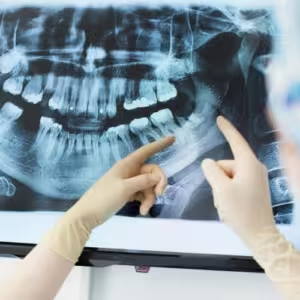
Dental X-rays, also known as radiographs, are essential diagnostic tools that play a crucial role in assessing oral health. They provide valuable insights that are not visible during a regular dental exam, helping dentists identify and treat dental problems early. At Dental 32 in Ashburn, VA, Dr. Ninh and his team utilize various types of dental X-rays tailored to meet the specific needs of adult patients. Let’s explore the different types of dental X-rays commonly used for adults:
1. Intraoral X-Rays
Intraoral X-rays are the most common type of dental X-rays and provide detailed images of the teeth, gums, and surrounding structures inside the mouth. These X-rays are particularly useful for detecting cavities, examining tooth roots, and assessing the health of the bone surrounding the teeth. There are several types of intraoral X-rays:
Bitewing X-rays: These X-rays show the upper and lower teeth in a specific area of the mouth. They are used to detect decay between teeth and assess the fit of dental restorations.
Periapical X-rays: Periapical X-rays capture the entire tooth from the crown to the root and the surrounding bone structure. They are used to diagnose dental issues such as abscesses, root infections, and bone loss.
Importance: Intraoral X-rays provide detailed information that helps dentists accurately diagnose and treat dental problems early, preventing complications and preserving oral health.
2. Panoramic X-Rays
Panoramic X-rays, also known as panoramic radiographs or panorex, provide a broad view of the entire mouth in a single image. This type of X-ray captures the teeth, jaws, sinuses, and surrounding structures. Panoramic X-rays are useful for:
Assessing Impacted Teeth: They help identify impacted wisdom teeth and other teeth that may not have erupted properly.
Evaluating Jaw Joint (TMJ) Issues: Panoramic X-rays can reveal issues affecting the jaw joints (temporomandibular joints) and surrounding bones.
Importance: Panoramic X-rays offer a comprehensive view of the oral and maxillofacial structures, aiding in the diagnosis of complex dental conditions and treatment planning.
3. Cone Beam Computed Tomography (CBCT)
CBCT is a specialized type of dental X-ray that provides three-dimensional (3D) images of the teeth, jawbone, nerves, and soft tissues. Unlike traditional X-rays, CBCT produces detailed cross-sectional views, offering precise information for:
Implant Planning: CBCT scans help dentists assess bone density and structure for precise placement of dental implants.
Orthodontic Treatment: They assist in planning orthodontic treatment by evaluating tooth and jaw relationships in 3D.
Importance: CBCT scans are invaluable for complex dental procedures, ensuring optimal treatment outcomes with minimal invasiveness and maximum precision.
4. Extraoral X-Rays
Extraoral X-rays are taken outside the mouth and focus on the jaw and skull. They are less detailed than intraoral X-rays but provide valuable information for:
Orthodontic Treatment: They help assess jaw development, tooth eruption patterns, and alignment issues.
Diagnosing Temporomandibular Joint Disorders (TMD): Extraoral X-rays can reveal abnormalities in the jaw joint and surrounding structures.
Importance: Extraoral X-rays complement intraoral X-rays by providing a broader perspective of dental and facial structures, aiding in comprehensive treatment planning and diagnosis of conditions affecting the jaw and skull.
5. Digital X-Rays vs. Traditional Film X-Rays
Modern dental practices, including Dental 32, utilize digital X-ray technology, which offers several advantages over traditional film X-rays:
Reduced Radiation Exposure: Digital X-rays emit significantly less radiation compared to traditional film X-rays, promoting patient safety.
Faster Results: Digital X-rays produce images instantly on a computer screen, eliminating the need for film processing and reducing appointment times.
Enhanced Image Quality: Digital X-rays can be manipulated and enhanced for better visualization of dental structures, aiding in diagnosis.
Importance: Digital X-ray technology enhances efficiency, improves diagnostic accuracy, and enhances patient comfort during dental exams.
Conclusion
Understanding the different types of dental X-rays for adults is essential for appreciating their role in maintaining optimal oral health. At Dental 32 in Ashburn, VA, Dr. Ninh and his team prioritize patient care and utilize state-of-the-art X-ray technology to provide accurate diagnoses and effective treatments. Whether you require routine X-rays during a dental check-up or specialized imaging for complex procedures, our commitment to advanced dental care ensures you receive the highest standard of treatment.
If you have questions about dental X-rays or would like to schedule an appointment at Dental 32, contact us today. Our knowledgeable team is here to provide comprehensive dental care tailored to your individual needs, ensuring a healthy smile and overall well-being. Remember, regular dental X-rays are a cornerstone of preventive care, helping you maintain a beautiful and healthy smile for years to come.
FAQ
Non-covered benefits may not be deemed medically necessary by insurance providers but can still be essential for maintaining dental health.
If a procedure isn’t covered by insurance, it’s essential to discuss alternative payment options with your dentist and budget for the expense accordingly.
Regular dental check-ups are critical for preventive care, regardless of insurance coverage. Skipping them can lead to more significant dental issues in the future
Budgeting for dental expenses ensures that you can cover the costs of non-covered benefits and access necessary treatments when needed.

